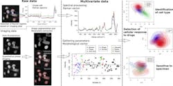
Researchers at Osaka University (Osaka, Japan) have developed a label-free multimodal imaging platform that enables the study of cell cultures noninvasively without the need of any contrast agent.
The two researchers—Assistant Professor Nicolas Pavillon and Associate Professor Nicholas I. Smith of the Immunology Frontier Research Center (IFReC) at Osaka University—showed how the label-free signals can be employed to create models that can detect the activation state of macrophage cells and distinguish between different cell types, even in the case of highly heterogeneous populations of primary cells.
"We devised specific statistical tools that allow for the identification of the best methods for detecting responses at the single-cell level, and show how these models can also identify different specimens, even within identical experimental conditions, allowing for the detection of outlier behaviors," says Smith.
The multimodal imaging platform they developed pairs quantitative phase microscopy and Raman spectroscopy, both of which are label-free techniques. In their paper that describes the work, they demonstrate the platform's ability to extract biomarkers based on cellular morphology and intracellular content. These approaches have been previously used to characterize specimens and identify cells from different origins—however, the measurement of finer features than the cell type with these techniques has proven to be challenging.
The researchers' study findings show that the approach enables the study of live samples without requiring contrast agents, and can also achieve high sensitivity at the single-cell level. "In particular," says Pavillon, "our results show that this method can identify different cell subtypes and their molecular changes during the immune response, as well as outlier behaviors between specimens."
Full details of the work appear in the journal Science Advances.
Source: EurekAlert press release
Got biophotonics-related news to share with us? Contact Lee Dubay, Associate Editor, BioOptics World
Get even more news like this delivered right to your inbox
