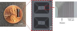
A team of scientists at Columbia University (New York, NY) used a microchip currently in development to map the back of the eye for disease diagnosis. So far, the scientists have been able to overcome the technical obstacles of current interference technology to fabricate a miniature device able to capture high-quality images.
In their paper that describes the work, the research team demonstrates the microchip's ability to produce high-contrast optical coherence tomography (OCT) images 0.6 mm deeper in human tissue.
Xingchen Ji, a lead author on the paper, hopes that the team receives additional industry funding to continue development of a small, fully integrated handheld OCT device for affordable deployment outside of a hospital in low-resource settings. The team has already received such funding from the National Institutes of Health (NIH; Bethesda, MD) and the U.S. Air Force (Washington, DC).
Central to chip-scale interferometry is fabrication of the tunable delay line. A delay line calculates how light waves interact, and by tuning to different optical paths, which are like different focal lengths on a camera, it collates the interference pattern to produce a high-contrast 3D image. Ji and co-author Aseema Mohanty coiled a 0.4 m silicon nitride (Si3N4) delay line into a compact 8 mm2 area and integrated the microchip with microheaters to optically tune the heat-sensitive Si3N4.
"By using the heaters, we achieve delay without any moving parts, so providing high stability, which is important for image quality of interference-based applications," says Ji.
But with components tightly bent in a small space, it's hard to avoid losses when changing the physical size of the optical path. Ji previously optimized fabrication to prevent optical loss. He applied this method alongside a new tapered region to accurately stitch lithographic patterns together—an essential step for achieving large systems. The team demonstrated the tunable delay line microchip on an existing commercial OCT system, showing that deeper depths could be probed while maintaining high-resolution images.
This technique should be applicable to all interference devices, and Mohanty and Ji are already starting to scale lidar systems, one of the biggest photonic interferometry systems.
Full details of the work appear in the journal APL Photonics.