"Without cytometric analyses, work in modern life sciences would be unthinkable." So begins chapter one in the text, Advanced Optical Flow Cytometry: Methods and Disease Diagnoses.1
Cytometry is the quantitative analysis of cells. As renowned cytometry expert Howard M. Shapiro explains in his book Practical Flow Cytometry (which Beckman Coulter has made available on its website for free as a searchable, printable, 733-page PDF file), it is a process designed to measure physical and/or chemical characteristics of single cells.2 It includes identifying, counting, and characterizing by multiple means, but most notably through the use of flow cytometers, which measure cells suspended in fluid as they zoom single-file past an illumination source.
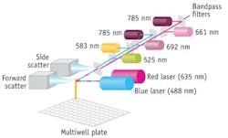
Recent trends in commercial flow cytometry instrumentation are size reduction, modularity, and ease of use for broader accessibility; increased availability of both wavelengths and targets; and incorporation of imaging capabilities. And more technical advances are on the horizon (see Fig. 1).
Industry trends are toward consolidation, as the few firms that have dominated this market continue to do so through acquisition. And according to Venture Planning Group's (New York, NY) "The 2012 Flow Cytometry Market" reports, advances in molecular diagnostics, monoclonal antibodies, lasers, and information technology—as well as growing understanding of immunologic forces regulating systemic diseases—will have a profound impact on the flow cytometry markets worldwide.
Hellma (Plainview, NY), maker of micro flow channels for cytometry systems, calls cytometry "one of the most powerful and important technologies for applied sciences in the 21st century."
Evidence in the market
Traditionally, the flow cytometry market has been dominated by a few large players, including Beckman Coulter (Brea, CA) and Becton, Dickinson and Co. (BD; Franklin Lakes, NJ). While a number of startups have launched to commercialize technology innovations, domination of the few continues, for the most part, as the stalwarts purchase the startups. Not surprisingly, then, the technology trends mentioned above are evident in acquisition announcements. For instance, Beckman Coulter's April 2012 release announcing its acquisition of automated systems and reagents manufacturer Blue Ocean Biomedical (Pembroke Pines, FL) focuses on ease of use and broader accessibility. According to the statement by Beckman Coulter Life Sciences, the move provides the company with "low-complexity analyzers that integrate automated sample preparation" in order to directly address "emerging flow cytometry market trends." Blue Ocean's technology takes a sample-in, result-out approach, and the company claims to offer the first true integration of sample prep, handling, analysis, and data management: "Essentially, the world's first load-and-go flow cytometers," according to Blue Ocean CEO and co-founder Mike Brochu.
Just about a year before, in February 2011, medical devices conglomorate and Beckman Coulter rival BD purchased Accuri Cytometers, Inc. (Ann Arbor, MI)—reportedly paying more than $200 million for the opportunity to expand BD's presence into "the emerging affordable personal flow cytometer space," and to expand the use of flow technology into disciplines like environmental studies that have not traditionally used flow cytometry.
Also in 2011 (August), EMD Millipore (Billerica, MA), the Life Science division of Merck KGaA of Germany, purchased Amnis Corp. (Seattle, WA), manufacturer of cell analysis systems that integrate flow cytometry and microscopy and address a range of applications (the Amnis website has a nice overview: https://www.amnis.com/applications.html). For instance, Amnis' high-resolution system, ImageStream, has found a niche in oncology for leukemia and lymphoma research, thanks to its ability to do several tasks at once: Characterize the nucleus:cytoplasm ratio, produce an accurate mitotic index, measure signal transduction, follow the uptake of drugs into cells, and measure apoptosis. Amnis' more compact and affordable bench-top system, FlowSight, mirrors the size-and-affordability vision of EMD Millipore's 2009 acquisition of the Guava flow cytometry product range. Guava Technologies (Hayward, CA) had developed a vision for enabling flow cytometry outside of a centralized lab and introduced bench-top systems using microcapillary technology, which accommodates relatively smaller sample volumes (see Fig. 2). Additionally, the company offered kits optimized for various research areas-stem cell, cancer biology, etc.—to save scientists from having to source reagents and develop their own assays.Demonstrating the trends of both modularity and more colors is the S1000Ex by Stratedigm (San Jose, CA), one of the innovators not yet purchased by a larger rival. This, Stratedigm's flagship flow cytometer, is equipped with up to four lasers to measure up to 18 parameters. It incorporates the company's Smart Detect architecture, which enables the use of a single integrated photomultiplier tube (PMT)/electronics module for detection of multiple fluorophores with similar emissions wavelengths, but different excitation lasers. According to Stratedigm CEO Shervin Javadi, this will enable multi-color analyzers capable of enhanced performance at lower prices than conventional products.
Components fuel progress
Suppliers to the instrumentation developers are working to facilitate these technology trends. Venture Planning Group notes that compact and easy-to-operate laser systems will further expand applications of flow cytometry to routine clinical laboratories. Toptica Photonics' iChrome is an example of this: The product won accolades upon its release for its compact size and ability to target multiple, specific fluorophores with automatically tunable narrow-band output.
In his 2011 article, "Seeing more detail with cytometry," BioOptics World contributing editor Mike May describes Omega Optical's (Brattleboro, VT) low-cost approach to enabling more wavelengths: Making multiple gratings along an optical fiber, each one of which can be adjusted to a particular center wavelength. He also explains the work of researchers at the University of California, Los Angeles, who were able to turbo-charge flow cytometry throughput with a 256-channel microfluidics device that hurried 28,000,000 cells through every second.
Beyond the usual suspects—that is, the light sources, optics, and flow channels—Venture Planning Group notes that influences on flow cytometry will come from other sources. For instance, information technology will eventually reduce the cost of instrument manufacture and service warranty, and permit development of self-troubleshooting, autocalibration, and other advanced features, such as automated analysis of chromosomal abnormalities, DNA content, and lymphocyte subsets. This will make routine procedures that today are still somewhat exotic, bringing the advantages of flow cytometry to more and more healthcare consumers.
In the meantime, the latest news reports progress toward portable systems for point-of-care applications: Researchers at the University of Toronto (Toronto, ON, Canada) have developed a flow cytometer specifically geared to track HIV progression in developing countries. While today's compact cytometers are often the size of a photocopier, this unit is the size of a bread loaf—and the team is planning to reduce it to fit in the palm. System cost is targeted at $5,000 to $10,000, with integrated wireless data communications, GPS, and camera. The team's goal is to have the handheld version ready for use in the field by mid-2012. And LeukoDx (Jerusalem, Israel) recently announced investment of up to $8 million over the next three years to enable further development of its own flow cytometry platform, which hopes to deliver test results in 10 minutes from a single drop of blood. The device promises to enable fast clinical decision-making in neonatal and intensive care units, emergency departments, and in remote locations.
REFERENCES
1. A. Mittag and A. Tárnok, Advanced Optical Flow Cytometry: Methods and Disease Diagnoses, John Wiley & Sons, Inc., Marblehead, MA (2011).
2. H.M. Shapiro, Practical Flow Cytometry, Wiley-Liss, 4th edition (2003).


