The Sutter BOB™ — designed to eliminate the conventional microscope frame—is a simple, open-design upright microscope platform ideal for slice electrophysiology, wide field functional imaging, two photon retinal imaging, photostimulation and new techniques just being developed! A microscope, in its simplest form, is an objective and a tube lens. Other components of most modern microscopes are designed to serve specific functions: different types of experiments, methods of illumination and means of signal detection.
Replacing the microscope frame with an optical rail builds in the ability to adjust the overall height of the microscope, unheard of in conventional microscope designs. Work on slices in January, do in vivo experiments in March. The BOB microscope is a compact, single assembly that mounts to the “blue rail” with one massive, stable connection. Focusing is motorized and incorporated between the focus arm and the optical rail.
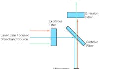
Fluorescence epi-illumination is built into the basic BOB via an Olympus vertical illuminator. LED transmitted light illumination uses the Olympus Oblique Coherent Contrast (OCC) condenser. Sutter’s TLED and TLED controller form the trans-illumination light source. The TLED controller is capable of being triggered with a digital signal eliminating the need for shutters and adding the ability to photostimulate from the trans location. In experiments where transmitted light is not desired, the LED, condenser focus mechanism and OCC condenser are easily removed as a single assembly. Additionally, the transmitted light path is shorter than in other frames, allowing the microscope body to sit significantly lower than a conventional microscope. When the microscope is shorter, there is more stability, increased ergometrics and ease of use.
The Sutter BOB when configured with an optional motorized XY stage or translator with MPC-200 controller, takes full advantage of our free Multi-Link™ software program for micromanipulator positioning. During whole-cell patch recording in slices, it is common to search a large area of the slice to find appropriate neurons. If the BOB is configured with Multi-Link, after you find your target, Multi-Link will then retrieve your recording and stimulation pipettes to the same field of view so that you can begin recording immediately. If later you need to stimulate a region outside the current field of view, Multi-Link can release the recording pipette and allow you to reposition the objective and stimulating pipette(s) to the new stimulation region.
- APPLICATIONS
- Patch clamp electrophysiology
- in vivo, in vitro, and slice
- Whole-cell recording
- Intracellular recording
- Material science
- FEATURES
- Optional motorized fixed XY stage or motorized translator
- Open design microscope with motorized focus
- Quickly configurable based on experimental needs
- Optimized to allow in vivo and in vitro experimentation on one setup
- Designed for use with Olympus objective lenses
- Free Multi-Link™ software coordinates movement with micropipette positioning of MPC-200
- Oblique Coherent Contrast (OCC) or Differential Interference Contrast (DIC)
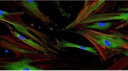
- Epi-fluorescent illumination