The Movable Objective Microscope® (MOM®) is a two- or thee-photon microscope capable of imaging deep within living specimens when combined with an appropriate laser. The Sutter MOM was the first scope to provide 3-dimensional objective movement and rotation allowing the specimen to remain horizontal and stationary. Many highly regarded imaging laboratories around the world use the Sutter MOM and we constantly work with our customers to adapt the design for their changing needs.
Watch the video describing MOM imaging and photostimulation beam paths
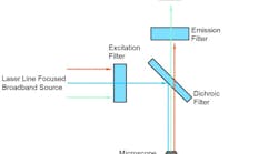
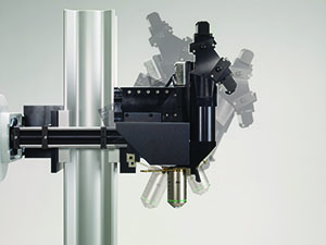
MOM Opto-mechanical DesignThe MOM consists of two independent microscopes. The wide-field half of the microscope consists of an Olympus vertical illuminator, Sutter Xenon arc lamp and camera mount to provide standard epifluorescence. The two-photon side of the microscope provides the optical pathway for guiding the excitation laser light from the table up into the scanning galvanometric mirrors and then expanding the beam through the scan lens and directing into the back of the objective. Following twophoton excitation, the emitted photons are directed by a dichroic mirror immediately above the objective into the detection pathway. The main body of the microscope moves backwards on a rail system allowing easy access to the specimen prior to imaging.
The objective translates in X, Y and Z as well as rotates around the X axis. Two moving mirrors allow the microscope to maintain efficient delivery of the excitation light to the back aperture of the objective regardless of movement or orientation. The X, Y and Z movements used are the same as that in our MP-285 micromanipulator so you know the movements are smooth, fine in scale, drift-free and highly reproducible. These movements permit Z-stacks and mosaic images of large regions of tissue to be recorded without the need for a moving stage.
The horizontal light path allows for rotation of the objective away from the standard vertical position. As a result of this rotation, the MOM can easily be converted from an upright to an inverted microscope and the objective positioned from 0 to 180 degrees. This positional freedom permits the imaging of non-horizontal surfaces and volumes.
MOM Scanning SystemsDuring the last 10 years, scanning systems for multiphoton microscopes have changed in several ways. Large aperture, high NA objectives became available and thus required larger aperture scanners. Resonant scanner technology allowed faster imaging. Two-photon scopes now include both resonant-galvo and resonant galvo-galvo systems. The Sutter MOM developed in parallel with these changes and new technology can be bolted into older, existing scopes with minimal changes. Many original scopes with 3 mm galvo scanners have been upgraded to either 6 mm galvo scanners or resonant/galvo scanners. As an example, the Vidrio RMR scanner (a resonant galvo-galvo scanner system) can be purchased as part of any new MOM system or retrofit into existing MOM scopes.
Imaging SoftwareStarting in 2011, Sutter began offering the MOM Computer System and Software (MCS). Before this software package was developed, most users relied on ScanImage or MPScope to generate scanned images. Customers valued the fact that the MOM would operate with open source freewares, however, there seemed to also be a market for a commercial package. MCS continues to offer a simple, easy to use package available at a price that compares with other commercial and freeware packages. MScan 3.0, the latest version, is Windows 10 compatible. A recent publication takes advantage of the long (1-2 hour) data files that can be captured in the MCS proprietary data file structure. (reference Kuhn, 2020).
The MOM® has always been compatible with ScanImage freeware, the two-photon imaging software developed by Karel Svoboda and collaborators. One of the reasons the MOM platform exists in its present form is the strong support from the ScanImage community. In 2014, Vidrio became the principle vehicle for support and new development of ScanImage. Sutter is happy to make Vidrio ScanImage Premium available to customers who wish premium support and the latest features. ScanImage Basic is available as an entry level system with a year of support included. ScanImage freeware is still available but does not include support. Sutter provides packages that include the necessary data acquisition hardware to couple the MOM and other scanning microscopes to ScanImage Premium, ScanImage Basic or the freeware version. We also sell Vidrio's hardware line including ScanImage ready computers, the vDAQ acquisiton system and the RMR scanner.
Sutter MOM packages include all of the equipment (less the laser and objective) needed for a complete imaging system:
- Scan lens and tube lens appropriate for two- or three-photon imaging
- Cambridge Technology XY galvonometric (3 mm or 6 mm) or resonant scanners (resonant-galvo or resonant galvo-galvo systems both with 5 mm mirrors)
- Hamamatsu photomultiplier tubes (PMTs): R6357 multialkali or H10770PA-40 (GaAsP) products. (Other PMTs are available, Sutter is an authorized reseller for Hamamatsu)
- Power supplies for PMTs: Sutter PS-2 (dual channel high-voltage power supply for conventional PMTs) or Sutter PS-2/LV (dual channel low-voltage power supply for H10770PA-40 or other PMTs with built in high voltage). Power supplies can be ordered with remote turn on/shut off for PMT gating
- Hamamatsu, Sigmann, or FEMTO pre-amplifiers
- Data acquisition: National Instruments PXI FPGA, Vidrio vDAQ, or National Instruments PC based Multifunction I/O
- Conoptics Pockels Cells for laser intensity control
- APPLICATIONS
- In vivo two-photon imaging
- In vivo three-photon imaging
- Electrophysiological recording and imaging (culture, large in vivo preparations, etc.)
- Non-horizontal surface microscopy
- Simultaneous retinal stimulation and two-photon microscopy1
- Whole animal imaging
- Immunology
- Embryology
- FEATURES
- Objective moves 22mm in X, Y, and Z
- Objective rotates about optical axis for imaging of non-horizontal surfaces and volumes
- Customizable open platform design
- Cambridge Technology conventional or resonant XY scanners
- Two or four channel detector system with Hamamatsu PMTs and preamplifiers
- Sutter PS-2/ PS-2LV dual channel PMT power supply
- Two- or three-photon compatible scan lens and tube lens
- *New* Light Block keeps visual stimuli, photostimuli, and ambient light out of detector path
- National Instruments and Vidrio Technologies data acquisition systems
1 "Eyecup scope-optical recordings of light stimulus-evoked flourescence signals in the retina", Euler et al, Pflugers Arch, 2008