OLIVER PUST
Good optical measurement is rarely, if ever, possible without high-performance optical filtering. Despite their critical role in all optical instruments, however, optical filters are often (and seemingly increasingly) considered only in the latter stages of instrumentation development—at which point system designers risk facing the unpleasant realization that an appropriate filter is unavailable and prohibitively expensive to design and produce.
Taking time to understand optical filters and considering them at the start of the design process has huge payoff potential for development of biomedical instruments able to dramatically shift patient care. It is also wise to select a filter manufacturer early on and involve them in the design process, as they can often contribute valuable insight to prevent wasted effort and unnecessary cost.
Novel cataract treatment
With an aging population, the clouding on the lens of the eye that causes degradation of vision—that is, cataracts—is a growing issue. Demand for cataract treatment is set to double by 2030. Ironically, the very cloudiness of a cataractous lens limits the ability to make diagnostic decisions. Researchers’ discovery that the fluorescence spectrum of the eye’s lens changes significantly as cataracts form, however, has led to a quantitative solution to the diagnostic problem.1 Fluorescence emission from the eye enables both early diagnosis and measurement of cataract development.
The current remedy for cataracts is a major surgery that involves extraction of the eye’s lens and insertion of a plastic implant. While the procedure is routine and highly successful, it demands highly trained surgeons and full operating theater resources. Opening of the capsule containing the natural lens (to insert the plastic lens) can induce opacity in the capsule—up to roughly 30% after three years, and 50% after five years. This is treated with another surgery (called capsulotomy) that involves use of a laser to create an aperture. But because some patients have fragile capsules, not all are good candidates for capsulotomy.
Edinburgh Biosciences has developed a method of cataract photobleaching that allows retention and preservation of the patient’s natural lens. To implement this process, the company has developed a compact instrument that facilitates not only tryptophan-based fluorescence detection for diagnosis and monitoring, but also a photobleaching (see Fig. 1). The closely monitored photobleaching process returns the lens’s fluorescence spectrum to match that of a healthy lens.
The monitoring module features a continuously variable bandpass filter (CVBPF) on a linear drive, and a single-photon counting photomultiplier tube (PMT) that collects the spectrum. Overall, the optical system—which also incorporates LEDs for both excitation and treatment, as well as phase-sensitive electronics—is 95% smaller than conventional instruments, and yet is still able to provide high light-gathering potential (light grasp) and operate in ambient light conditions.
Filter-expanded PCR
With its goal to transfer medical diagnostic and treatment capabilities from large, centralized laboratories and operation theaters to local clinics, inpatient beds, and even homes, point-of-care (PoC) instrumentation is a hot area of research and development. Currently, blood and other bodily fluids are routinely sampled in a healthcare provider’s office and sent to a central laboratory for analysis—a process that can delay treatment and create distress in patients. PoC instrumentation promises not only to alleviate the delay and distress, but also to bring advanced diagnostics to patients in remote locations and developing areas.
For practical implementation, PoC instruments must be compact, portable, and robust. Most of PoC technology’s potential applications require performance at least as capable as existing methods—for instance, equivalent sensitivity. Thus, all optical components must be reduced in size for PoC instruments, without compromising optical output. A related capability is collection of sufficient signal, which can impact filter performance. Moreover, small PoC instruments that are based on fluorescence signal detection typically provide insufficient space to accommodate the dichroic beamsplitter (which can help suppress excitation light in the emission channel) often used to separate excitation and emission channels. This increases the blocking requirements of the bandpass filters.
Cepheid’s GeneXpert Omni provides a successful example of device miniaturization, and transfer of diagnostic capabilities previously available only through full-size instruments. GeneXpert Omni provides portable clinical polymerase chain reaction (PCR) testing for molecular diagnostics using cartridge technology. Microfluidics in the cartridge regulate all aspects of the process, including sample preparation, extraction of the nucleic acid, amplification, and fluorescence-based detection. Custom-designed bandpass filters enable the instrument to perform many tests in addition to the HIV viral load test for which it was originally designed.2
Wavelength-selective biospectroscopy
In benchtop spectroscopic instruments, optical filters—often positioned on sliders or filter wheels—serve as wavelength selective elements in excitation and emission channels. Given that they have significantly greater light-gathering potential than gratings, filters convey important advantages. However, an instrument can accommodate only so many filters—a fact that restricts flexibility by making signal optimization difficult and time consuming.
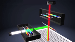
A new generation of multimode microplate readers by BMG Labtech uses continuously variable filters (CVFs) to provide high light grasp (and thus high sensitivity), the tunability of a grating-based instrument, and both narrow and wide passbands for stronger signal collection. A continuously variable dichroic (CVD) mirror allows the instrument to separate excitation and emission light in an epifluorescence setup (see Fig. 2).Besides their blocking capabilities, modern CVBPFs feature passbands narrow and steep enough for many spectroscopic applications (for example, fluorescence or color measurements) and high transmission levels. Their diminutive size allows them to be combined with typical line or array sensors—to enable point measurements, or novel multi- or hyperspectral camera design, respectively.
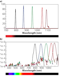
Wavelength resolution is a key parameter of any spectrometer. In the case of diffractive spectrometers, it is determined mainly by the size of the slit; sub-nanometer resolution can be achieved in exchange for optical throughput. For spectrometers based on CVBPFs, spectral resolution is a combination of factors—most importantly the bandwidth of the CVBPF. This theoretical value is defined for collimated light illuminating an infinitely small spot on the filter, which is the result of the bandpass layer structure. A multicavity design, naturally exhibiting narrow bands and steep edges, is typical. The bandwidth is in direct proportion to the center wavelength in a first approximation.
When a CVBPF is paired with a pixel-based sensor, spectral performance corresponds not to the spectrometer’s slit size, but to the sensor’s pixel size, which is typically a few micrometers. Practically speaking, this means that a single pixel receives light with spectral width correlated to the CVBPF’s bandwidth—normally several nanometers. Spectral resolution, then, is determined by a combination of filter bandwidth, filter wavelength gradient, and the pixel pitch of the sensor.
For more detail on the use of optical filters for spectroscopy, please see Portable Spectroscopy and Spectrometry 1: Vibrational, Optical and Bioanalytical Instrumentation and Applications, edited by Richard Crocombe, Pauline Leary, and Brooke Kammrath, by Wiley Publishing (2018).
REFERENCES
1. D. M. Gakamsky, B. Dhillon, J. Babraj, M. Shelton, and S. D. Smith, J. R. Soc. Interface, 8, 64 (2011); http://doi.org/10.1098/rsif.2010.0608.
2. See http://bit.ly/Cepheid.
Oliver Pust is Director, Sales and Marketing for Delta Optical Thin Film A/S, Hørsholm, Denmark; e-mail: [email protected]; www.deltaopticalthinfilm.com.
![FIGURE 1. This functional prototype of a combined system for diagnosing, monitoring, and treating cataracts includes eye positioning location (a), LEDs to excite fluorescence for quantitative diagnosis and to provide photobleaching treatment (b), a continuously variable bandpass filter (CVBPF)-enabled spectrometer (c), a single-photon counting photomultiplier tube (PMT; [d]), and a second eye-positioning location (e). FIGURE 1. This functional prototype of a combined system for diagnosing, monitoring, and treating cataracts includes eye positioning location (a), LEDs to excite fluorescence for quantitative diagnosis and to provide photobleaching treatment (b), a continuously variable bandpass filter (CVBPF)-enabled spectrometer (c), a single-photon counting photomultiplier tube (PMT; [d]), and a second eye-positioning location (e).](https://img.laserfocusworld.com/files/base/ebm/lfw/image/2020/03/2004LFW_pus_1.5e8346083bae0.png?auto=format,compress&fit=max&q=45&w=250&width=250)

