Biomedical researchers and optical engineers have long used modeling software to streamline the development of new products and applications. Now, Breault Research Organization (BRO; Tucson, AZ) is augmenting the optical-modeling capabilities of its optical-design software, the Advanced Systems Analysis Program (ASAP), with a skin-modeling technique called Realistic Skin Model (RSM).
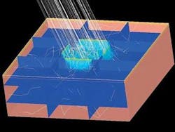
According to Breault, the RSM is capable of modeling the major layers of human skin—stratum corneum, epidermis, dermis, and hypodermis—independently (see figure). This approach allows layer-by-layer customization of models, providing a tissue model tailored to precise specifications. Built into the RSM are options for adjusting concentrations of light-absorbing molecules, or chromophores, in each skin layer. This feature opens up an array of possibilities for disease and disorder modeling. The RSM tissue models can be integrated with pre-existing optical designs for accurate characterization of system functionality and optical phenomena in tissue samples. Simultaneous modeling of optical systems and tissue makes virtual analysis of the light-tissue interface a fast and low-cost alternative to laboratory studies.
Paul Holcomb, a BRO biomedical engineer who developed the RSM, notes that modeling optical and biological components at the same time is important for designing surgical and other medical instruments. “You can test them on realistic skin models to see, for instance, what happens when you put a certain laser energy into a tissue sample, and where the energy goes,” he says.
Phantoms in the framework
So-called tissue phantoms can be created in ASAP and placed within a modeled optical system, allowing for the characterization of system performance within a biologically accurate framework. Using a scatter and absorption model that relies on nonsequential ray-trace analysis, the RSM replicates the absorption and scattering properties of human skin. The major chromophores of skin—melanin, hemoglobin, and water—are represented within the model, along with lesser contributors such as bilirubin and beta carotene. The absorption and scatter characteristics of each layer of tissue are calculated independently, taking into account the different chromophore concentrations in each layer.
“The skin is one of the most complex and dynamic organs of the human body. It can be weathered by age, transformed by disease, and marred by genetics,” Holcomb said. “Modeling of the skin in any accurate manner is necessarily complex, and no model can completely encompass all factors involved. The RSM takes into account as many biologically relevant aspects of the human skin as is feasible.”
The program uses the Henyey-Greenstein model. Four parameters are required to create this model: the anisotropic scatter coefficient, the scattering coefficient, the absorption coefficient, and the fractional obscuration per unit area. Each of these factors is wavelength-dependent. Concentrations of every component used in the modeling process can be controlled, allowing many different skin conditions and disorders to be modeled. A single tissue phantom can be created and placed within an existing optical system, or the entire trace and analysis process can be accomplished from the RSM dialogs.
“The amount of oxy- and deoxy-hemoglobin can be adjusted to examine the effects of oxygen deprivation on skin optical properties, or the amount of bilirubin can be increased to determine the effects of jaundice, a common symptom of liver disease,” Holcomb said.
Ultimately, BRO plans to build a library of models similar to the RSM that would contain not only the absorption and scattering of tissues, but also realistic optical properties of internal organs such as the bladder.
