Benjamin Potsaid, James Jiang, Vijaysekhar Jayaraman, Alex Cable, and James G. Fujimoto
Optical coherence tomography (OCT) is commonly thought of as a millimeter-range imaging technology. With the recent introduction of new OCT light sources based on vertical-cavity surface-emitting laser (VCSEL) technology, by researchers at Thorlabs, Praevium Research, and the Massachusetts Institute of Technology (MIT), this understanding may be about to change.1 VCSEL-based OCT imaging achieves a combination of long imaging range, high speed, and flexibility in operation that potentially opens the door for new applications in biomedicine—as well as other areas.
OCT imaging works like an optical version of ultrasound but instead of sound, infrared (IR) light is used to probe and visualize the structure of tissue and organs. While ultrasound produces image resolution on the scale of a tenth of a millimeter, OCT can achieve orders-of-magnitude finer resolution on the micrometer scale.
In OCT, the axial resolution is related to the bandwidth of light used in the interrogation: The wider the span of wavelengths in the light, the finer the axial resolution. Using a technique called swept-source OCT, the microelectromechanical systems (MEMS)-based tunable VCSEL developed by the researchers achieves fine resolution by repetitively sweeping the output wavelength of light over a broad spectrum in time. It is the manner in which the light is swept that differentiates the VCSEL from previous swept-source OCT technologies.
VCSEL structure and operation
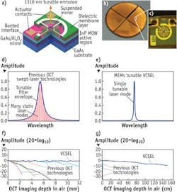
The VCSEL is fabricated using semiconductor processing techniques (see Fig. 1). A laser cavity is formed by locating a gain material between two mirrors, one of which is stationary and the other of which is suspended by a flexible structure. The mirrors form a Fabry-Perot filter such that the wavelength of tuned emission is proportional to the distance separating them. With the application of voltage, an electrostatic MEMS actuator pulls the top mirror down, thereby reducing the cavity length and tuning a shorter wavelength of emission. It is the monolithic design and micron-scale cavity length that enable the VCSEL to produce true single-mode and mode-hop free tuning.
Previously demonstrated OCT swept light sources consisted of a relatively long laser external cavity ranging from a few centimeters to a few meters in length. Tuning was accomplished through a tunable intracavity filter or tunable wavelength selective mirror, which necessarily selected a cluster of modes and provided multimode tuned operation. Shortening the cavity in these designs ends up selecting fewer modes, but introduces discontinuous mode-hopped operation. The VCSEL provides a new operating regime in which a few-microns-long Fabry-Perot cavity comprises the entire laser cavity, pushing the free spectral range (FSR) beyond 100 nm and enabling mode-hop-free single-mode tuning over this entire FSR. The result is that the VCSEL has a coherence length much longer than has ever been seen in OCT swept light source technologies, which enables the unique long OCT imaging range of the VCSEL.2
Long-range imaging
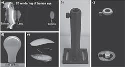
Because current generation OCT technologies are limited to a few millimeters of range, ophthalmic clinics must use three separate OCT instruments for 1) imaging the retina, 2) imaging the anterior eye, and 3) performing axial eye length measurements. A recent paper demonstrates the unique long-range OCT imaging capability of the VCSEL, which enables, for the first time, simultaneous imaging of the anterior eye and measurement of the full length of the eye from the cornea to the retina in a single OCT acquisition (see Fig. 2).3 This constitutes a first step towards an integrated three-in-one ophthalmic instrument able to save cost and space, and streamline patient flow in the clinic.This capability to image over longer ranges is promising for many non-medical applications, too. Recent demonstrations include non-destructive imaging inside a light bulb and 15.2 cm imaging of an optical post with precise depth measurement inside a high-aspect-ratio bore hole. Fast 3D OCT imaging of large objects such as these was inconceivable with previous OCT technologies because of limited depth range. The ability to rapidly and non-destructively image in 3D and surface profile over tens of centimeters at high resolution promises to aid in manufacturing processes and quality assurance.
High-speed capture
The current generation of commercial OCT imaging systems operates at between 25 and 100 kHz axial scans rate. The short laser cavity and fast MEMS actuator in the VCSEL enable imaging at speeds up to 1.2 MHz axial scans rate—about 10–50x faster than current technology.
Applications that benefit from high-speed imaging include those involving movement or the need to image a large area. For instance, imaging of the human retina at a 1.2 MHz axial scan rate enables comprehensive coverage of the retina with high lateral pixel density (see Fig. 3). A retinal scan obtained at a 400 kHz axial scan rate can be motion-corrected and averaged from four volumetric acquisitions. For cancer imaging and diagnostics, the high speeds are critical for comprehensive coverage of large areas and to achieve high-lateral-resolution imaging. Using a miniature endoscopic probe, MIT researchers and clinical collaborators produced in vivo images of a rabbit stomach using a 1 MHz axial scan rate as a translational step towards human endoscopic demonstrations.4Precise dynamics measurement
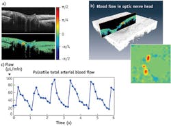
Doppler OCT allows the velocity of moving particles in a sample to be measured in the direction of the incident light beam. Precise measurement of the dynamics of the blood flow in the human eye may improve early detection of eye disease. In the eye, the blood enters and exits through the optic nerve head. The high imaging speeds and good phase stability enabled by the VCSEL allow for measurement of rapid blood flow in the optical nerve head (see Fig. 4). Further, high speeds facilitate the collection of multiple repeated 3D volumes to generate a comprehensive time history of blood flow into and out of the eye.5 Plotting the data from each volume, it is possible to observe pulsatility within the eye.Phase-based OCT methods such as Doppler in general are much more sensitive to movement than intensity methods, and have been used to measure picometer displacements and micro-resonances in structures. The VCSEL may be beneficial for measuring dynamic phenomena, such as shock waves traveling through materials or dynamic movements of micro and macro structures.
While MIT and Praevium are in the process of deploying VCSEL-based prototypes to clinics with support from the National Institutes of Health Cancer and Eye Institutes (NCI grant R44CA101067 and NEI grant 1R44EY022864-01), Thorlabs is commercializing the MEMS-VCSEL technology to be offered as a complete OCT imaging system, as a benchtop laser, or as a swept source for OEM system integration.
REFERENCES
1. V. Jayaraman et al., Conference on Lasers and Electro-Optics (CLEO), PDPB2 (2011).
2. B. Potsaid et al., "MEMS tunable VCSEL light source for ultrahigh speed 60 kHz-1 MHz axial scan rate and long range centimeter class OCT imaging," Proc. SPIE, 8213, 82130M-8 (2012).
3. I. Grulkowski et al., Opt. Lett., 38, 673–675 (2013).
4. T-H Tsai et al., "Ultrahigh speed endoscopic optical coherence tomography using micro-motor imaging catheter and VCSEL technology" Proc. SPIE, 8571, 85710N (2013).
5. W. Choi et al., Opt. Lett., 38, 338–340 (2013).
Benjamin Potsaid, PhD, is a research scientist at Thorlabs (Newton, NJ) and a visiting scientist at the Research Laboratory of Electronics at MIT (Cambridge, MA); James Jiang, PhD, is the head of OCT research at Thorlabs; Vijaysekhar Jayaraman, PhD, is founder of Praevium Research (Santa Barbara, CA); Alex Cable is founder and president of Thorlabs; and James G. Fujimoto, PhD, is professor of electrical engineering at MIT. Contact Dr. Potsaid at [email protected].


