A research team at the University of Tsukuba (Tsukuba, Japan), led by associate professor Hideaki Kano, performed ultra-multiplex spectroscopic imaging (18 colors corresponding to 18 wavenumbers; 600–3600 cm-1) using coherent anti-Stokes Raman scattering on live A549 cells, reporting an effective exposure time per pixel of 1.8 ms.
Coherent anti-Stokes Raman scattering (CARS) allows unlabeled molecules in the human body to be identified via spectral analysis. Ultra-multiplex CARS is broad enough to detect all vibrational fundamental modes, including the critically important fingerprint region (in which unknown organic compounds can be identified and distinguished from one another) as well as the C-H and O-H stretching regions (useful for identifying the rough molecular distribution of lipids, proteins, and nucleic acids). Kano explains that the combination of this real-time, noninvasive, label-free technique and multivariate analysis methods can help life scientists and medical doctors gain meaningful insights into the dynamics of intracellular metabolic activity.
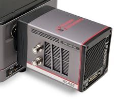
For their experiments, Kano's research group used a spectroscopy CCD camera (Teledyne Princeton Instruments' BLAZE camera) with super-deep-depletion CCD to yield high near-infrared quantum efficiency. The camera's sensor platform has dual 16 MHz readout ports to enable spectral rates of more than 1600 spectra per second with full vertical binning and up to 215 kHz when operated in kinetics mode. Its back-illuminated 1340 × 400 spectroscopic array format (20 μm square pixels) is capable of being cooled down to -95°C in air, without chillers or liquid assist, for low-dark-current performance. The researchers also used the company's LS-785 spectrograph and LightField software in their work.
Currently, the research group is collaborating with a pathologist to develop a new spectroscopic diagnostic tool using the molecular fingerprint.
Full details of the work appear in the journal OSA Continuum.
