After a little focusing up and down in a sample or cutting one thin section after another, every biologist wishes for one thing: deeper imaging. Ongoing advances in clearing agents, which make a sample more transparent, and microscope objectives that focus deeper into a sample give life scientists more information about biology's structure. In addition, combining these tools with labeling adds molecular information to the picture, and innovations in light sources enable deeper penetration through tissue. In this article, experts from Carl Zeiss Microscopy (Oberkochen, Germany), Leica Microsystems (Mannheim, Germany), and Olympus America (Center Valley, PA) reveal some of the advances in deeper microscopic imaging.
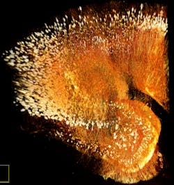
Although Brendan Brinkman, senior product manager at Olympus America, points out the broad interest among life scientists in seeing structures deeper below a sample's surface, he adds that "lots of the advancements in the technology have come in the brain sciences." The continuing interest in unraveling the connections in the brain keeps scientists on the lookout for new ways to map large volumes in a contiguous fashion (see Fig. 1).
Clearing the view
"One hot area is clearing agents," says Brinkman. "The brain is relatively opaque, but if you clear it and use confocal or multiphoton microscopy, you can see more deeply into the tissue."
For example, scientists at the RIKEN Brain Science Institute in Japan developed a clearing agent called Scale. Olympus offers this agent as SCALEVIEW, and the company has developed objectives optimized for this agent in 4 mm and 8 mm focal lengths (see Fig. 2). Other Japanese scientists developed the SeeBD clearing agent, while Karl Deisseroth and his colleagues at Stanford University developed CLARITY. Brinkman says, "Now, we also have an 8 mm objective that matches the refractive index of SeeBD and CLARITY." He notes that such lenses require many characteristics, including a high numerical aperture, matching the refractive index of the clearing agent, and collecting as many photons as possible. "We also add a correction collar on the lens to adjust for the thickness of the tissue," Brinkman says.To make such a system as easy to use as possible, Brinkman says, "Ours are as automated as they can be." This includes capabilities like "automatically increasing the laser power as you go deeper in the sample," he explains. "Also, if you change the objective, you can just tell the system, and it automatically adjusts laser properties to match."
The Leica IRAPO objectives from Leica Microsystems are also optimized for deep-tissue imaging with multiphoton microscopy. They are color-corrected for excitation up to 1300 nm and offer transmission higher than 85 percent for 470–1200 nm. "This technology allows efficient photon collection in deeper layers of tissues and protects precious samples since it requires less laser power," says Petra Haas, product manager at Leica Microsystems.
Also, Olympus recently updated its 25x lenses to a wide-band coating that allows for excitation from 400 to 1600 nm.
Increasing excitation
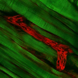
Other ways also exist for deeper imaging (see Fig. 3). "A typical multiphoton system can only go to excitation of 1040 nm," says Joseph Huff, product marketing manager for laser-scanning and super-resolution microscopy at Carl Zeiss. "Our Optical Parametric Oscillator, OPO, lets you get to 1300 nm. The longer wavelength provides deeper imaging, plus OPO lets you simultaneously use green and red fluorescent protein." Furthermore, Olympus America supports the InSight DeepSee ultrafast laser from Spectra-Physics that images out to 1300 nm.This system can show even more structures without labeling. For example, Huff says that second-harmonic excitation highlights muscle tissue, and the third harmonic emphasizes the myelin sheath around motor neurons. Huff adds, "OPO works on any multiphoton system that we offer."
With the Leica TCS SP8 MP microscope, researchers can also make use of the OPO lasers to get up to 1300 nm. In addition, Leica also offers the InSight DeepSee ultrafast laser. "To improve the user experience, the InSight laser is fully controlled by the LAS AF microscope software, like all other Leica multiphoton solutions," says Haas.
Deeper imaging also requires the right detector. The super-sensititve Leica HyD non-descanned detector is one option. According to Haas, the Leica TCS SP8 MP microscopes can soon be equipped with up to four of these detectors to simultaneously collect photons of different wavelengths.
The speed of the Leica TCS SP8, which scans at up to 428 frames/s at 512 × 16 pixels, and the high-speed z-control by the Leica GalvoFlow also benefit deep-tissue imaging. In combination, these technologies significantly reduce the time to acquire large image stacks.
Looking inside living samples
More and more, life scientists want to image intact living organisms—but depth becomes a real issue here. One solution, from Carl Zeiss, is the Lightsheet Z.1, which provides fluorescent imaging at depth in such living samples. While other fluorescent systems use the same light path for illumination and detection, the Lightsheet uses two separate paths. "The part of the tissue you are exciting is what you collect light from," says Scott Olenych, product marketing manager for imaging products at Carl Zeiss, "so there is no need for confocal or optical sectioning." In addition, this technique produces less photodamage, so researchers can do longer bouts of live imaging.
This technique also gives more options for angles on the sample. In essence, the sample gets suspended. "The objective is on the side with the sample hanging in front of it," Olenych explains. "You can rotate the sample and even choose the angle that is optimal for imaging." As a result, the resolution in the z-axis comes closer than usual to that of the x- and y-axes. To see even better, a researcher can combine a clearing agent with Lightsheet. For example, Lightsheet Z.1 has been used successfully with Scale.
These new tools—from clearing agents to objectives and complete systems—dramatically change how a biologist explores microscopic structures. Now, you can see deeper and in more colors, all while looking through tissue that, at times, appears almost transparent. When it comes to deeper imaging, it's a new world: one where we see so much more—more than we ever imagined.


